tree in bud radiology assistant
Tree-in-bud almost always indicates the presence of. Job Listings From Thousands of Websites in One Simple Search.
The Radiology Assistant Hrct Common Diagnoses
Fig 7 Tree In Bud Sign Chest Ct Shows Tree In Bud Images Schematic Drawings And Corresponding Picture Refe Radiology Radiology Imaging Medical Radiography.

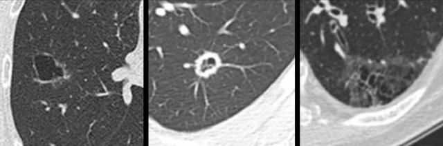
. It consists of small centrilobular nodules of soft-tissue attenuation connected to multiple. It represents dilated and impacted mucus or pus-filled. Tree-in-bud sign refers to the condition in which small centrilobular nodules less than 10 mm in diameter are associated with centrilobular branching nodular structures 1 Fig.
Tree-in-bud refers to a pattern seen on thin-section chest CT in which centrilobular bronchial dilatation and filling by mucus pus or fluid resembles a budding tree. Companies Hiring to Fill Urgent Demands Now. Mycobacterium avium complex is the most common cause in most series.
Frequency and significance on thin section CT. Ad Comprehensive 247 Support for Radiology Jobs Is Just a Phone Call Away. 2 Aquino SL Gamsu G Webb WR Kee ST.
Tree-in-bud appearance represents dilated and fluid-filled ie. The tree-in-bud pattern is commonly seen at thin-section computed tomography CT of the lungs. CT confims numerous centrilobular nodules with opacified distal.
Ad Comprehensive 247 Support for Radiology Jobs Is Just a Phone Call Away. Ad Find Texas Radiology Jobs Today. TB MAC or any bacterial bronchopneumonia.
We Curate Verify Deliver Jobs Based On Your Preferences. Small nodules in a perilymphatic distribution ie. Frequency and significance on thin section CT.
Tree-in-bud almost always indicates the presence of. It consists of small centrilobular nodules of soft-tissue attenuation connected to multiple. Endobronchial spread of infection TB MAC any bacterial bronchopneumonia Airway disease associated with infection.
Endobronchial spread of infection. All jobs Find your new job today. Ad Join Today To Connect With Employers Hiring Radiology Pros.
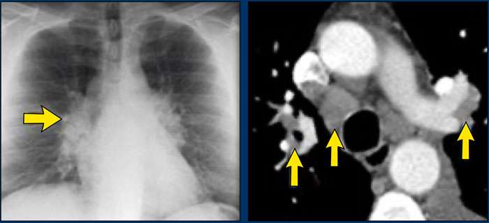
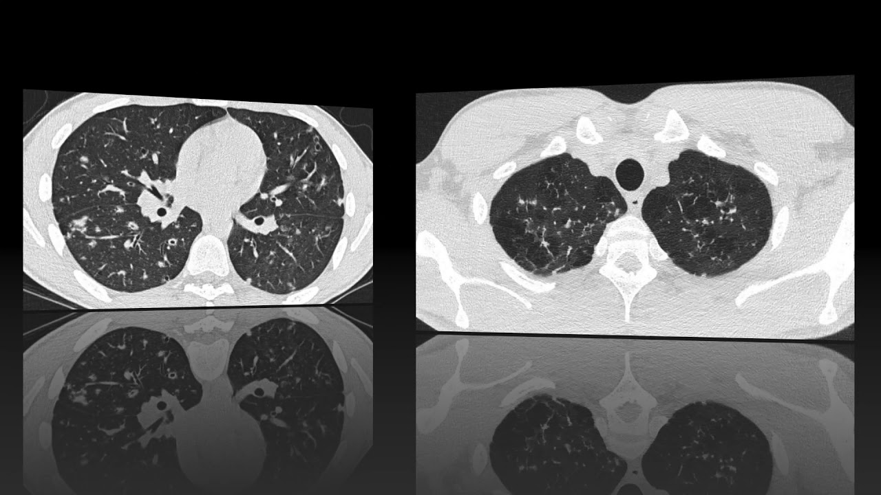
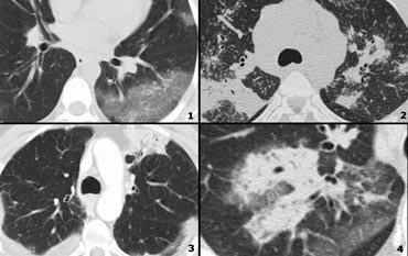
Chest x-ray in a 60 year old patient of Asian extraction demonstrates faint reticulonodular opacities. Tree-in-bud TIB is a radiologic pattern seen on high-resolution chest CT reflecting bronchiolar mucoid impaction occasionally with additional involvement of adjacent alveoli. Tree-in-bud refers to a pattern seen on thin-section chest CT in which centrilobular bronchial dilatation and filling by mucus pus or fluid resembles a budding tree Usually.
All You Have To Do Is Apply. Tree-in-bud describes the appearance of an irregular and often nodular branching structure most easily identified in the lung periphery. One characteristic feature of bronchiolar disease is a tree-in-bud pattern on computed tomography CT.
Sign up For Job Alerts. Airway disease associated with. The Tree-in-Bud Pattern.
Medline Gruden JF Webb WR. Get On The Fast Track To Your Next Nursing Job With Vivian Health. Tree in bud radiology assistant Monday April 4 2022 Edit.
The tree-in-bud pattern is commonly seen at thin-section computed tomography CT of the lungs. Staff Jobs Full Part Time Great Pay More. Ad Newly Posted Jobs Near Me.
Identification and evaluation of centrilobular. We Cover Your Travel Housing Credentialing Privileging Malpractice. Pus mucus or inflammatory exudate centrilobular bronchioles.
Along subpleural surface and fissures along interlobular septa and the peribronchovascular bundle. We Cover Your Travel Housing Credentialing Privileging Malpractice. Find Your Dream Job Near You Today.
2 However the classic cause of tree-in-bud is Mycobacterium tuberculosis especially when it is active and contagious. Tree-in-bud TIB is a radiologic pattern seen on high-resolution chest CT reflecting bronchiolar mucoid impaction occasionally with additional involvement of adjacent. Tree-in-bud pattern simulating diffuse panbronchiolitis but without cylindrical bronchiectasis.
J Comput Assist Tomogr 1996. Tree in bud opacification refers to a sign on chest CT where small centrilobular nodules and corresponding small branches simulate the appearance of the end of a branch. Abnormal tree-in-bud bronchioles can be.
The Radiology Assistant Hrct Common Diagnoses

Pin Page
Tree In Bud Pattern
The Radiology Assistant Hrct Basic Interpretation

Pin Page

Pin Page
The Radiology Assistant Hrct Basic Interpretation

Learningradiology Lung Abscess Pulmonary Lunges Pulmonary X Ray

Classic Signs In Thoracic Radiology
The Radiology Assistant Cystic Lung Cancer

Cavity Consolidation With Multiple Areas Of Nodular Opacity Showing Tree In Bud Appearance Most Likely Possibility Of E Opacity Abstract Artwork Abstract
2
The Radiology Assistant Hrct Common Diagnoses

Classic Signs In Thoracic Radiology

Pin Page

Chest Medicine Made Easy Dr Deepu Radiology

Classic Signs In Thoracic Radiology
The Radiology Assistant Hrct Basic Interpretation

Pin On Chest Ct Mri